David E. Marrus Adaptive Optics Imaging Laboratory at NYEE
“If we’re hoping to save sight by initiating earlier treatment, we need high-resolution pictures to detect the most subtle changes in the eye,” explains Toco Chui, PhD, Director of the Marrus Adaptive Optics Laboratory and Associate Professor of Ophthalmology at the Icahn School of Medicine at Mount Sinai.
One of the gems of the Einhorn Center is the Marrus Adaptive Optics Imaging Laboratory, which is advancing the science of ocular imaging to the next level. Our Adaptive Optics Imaging Laboratory is one of a handful of sites in the country with the Milwaukee Adaptive Optics Scanning Light Ophthalmoscope (AOSLO), a sophisticated imaging system that delivers unprecedented visualization of the retinal microvascular structure and blood flow. NYEE’s researchers have helped develop analytical software to harness AO’s ability to reveal microanatomical features of the retina in living patients.
Imaging Plays a Crucial Role in Patient Care
NYEE has been a pioneer in early eye disease detection with a long history of developing and refining imaging tools that access the microstructures of the eye, such as optical coherence tomography (OCT), scanning laser ophthalmoscopy (SLO), high resolution ultrasound, retinal blood flow imaging, macular pigment densitometry, retinal oximetry, and metabolic mapping. The addition of adaptive optics SLO (AOSLO) imaging, which enables acquisition of high-resolution cellular information in living patients, has dramatically changed the way vision scientists and ophthalmologists investigate the retina, and ultimately the way eye diseases are diagnosed and treated. The enhanced views of the eye and its structures help NYEE physicians make earlier, more accurate diagnoses, as well as monitor cellular effects of therapeutic interventions.
Going Deeper With Adaptive Optics
With advances in pharmacological therapies and biological treatments such as stem cells and gene therapy, the need for techniques to detect earlier changes in patients’ eyes became apparent. Adaptive optics, with its high-resolution and non-invasive advantages, is well suited for retinal imaging in living patients, and allows direct visualization of individual rod and cone photoreceptor cells, capillaries, and nerve fibers. The technology was introduced into astronomical telescopes by the U.S. government nearly 30 years ago to improve image resolution by compensating for distortions caused by atmospheric turbulence. In the case of ocular applications, adaptive optics helps achieve cellular-level imaging by overcoming the aberrations to light illuminating the eye. This is accomplished by measuring the distortions of the wavefront of the light and correcting them using a special “adaptive” mirror that essentially eliminates the distortions. Adaptive optics improves the optical transmission of light to lateral resolution to better than 2 microns, 10 times that of OCT imaging. This is sufficient to image and count the individual rods in the photoreceptor mosaic, which are approximately 2.5 microns in diameter.
Taking Adaptive Optics (AO) to the Next Level
Building on the sophisticated imaging capabilities of adaptive optics, the researchers at NYEE have made it an even more powerful tool by developing software for measuring features revealed in these images. These analytic tools have helped clinicians convert the reams of data gathered into early signals of disease, which can be used to monitor progression or response to treatment.
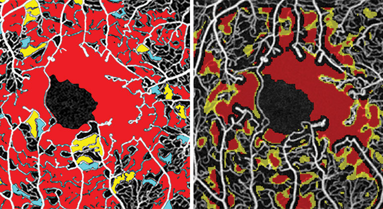
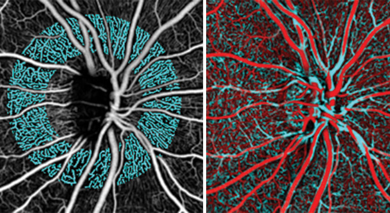
- Adaptive Optics + OCTA: At NYEE, adaptive optics has been paired with optical coherence tomography angiography (OCTA) to help transform the early detection of primary open-angle glaucoma and normal-tension glaucoma. To that end, research at NYEE has identified subtle changes in the retinal nerve fiber layer and its supportive radial peripapillary capillary (RPC) network.
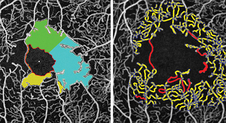
- AOSLO: NYEE’s researchers found that microscopic, non-invasive imaging of the fine retinal vasculature using non-confocal AOSLO enabled quantification of lumen diameter and wall thickness. Significantly, they learned this technique has potential for earlier detection of diabetic retinopathy, as well as more accurate monitoring of its progression and any treatment response.
- Adaptive Optics-OCT: NYEE researchers are currently developing plans for a multi-institutional collaboration to add adaptive optics OCT instrumentation to our current instrumentation suite. This will provide enhanced choroidal imaging for a series of upcoming macular degeneration and optic nerve studies.