A Leader in Specialty Eye Care
Ranked as one of U.S. News & World Report's® “High-Performing” Hospitals for Ophthalmology for 2023-24
Eye Services
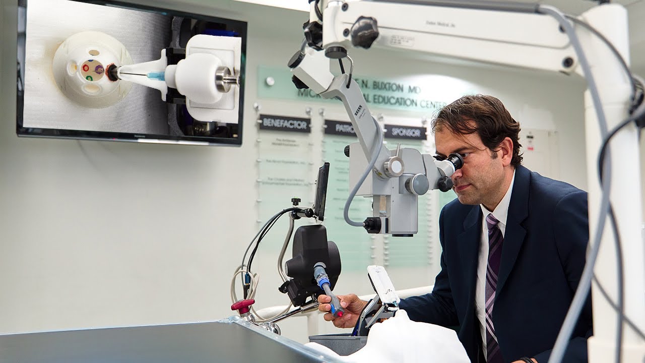
New York Eye and Ear Infirmary of Mount Sinai (NYEE) offers patients the most advanced and comprehensive treatments for all eye conditions. Our highly skilled physicians are experts in managing the full range of eye problems, including cataracts, corneal disease, eye trauma, glaucoma, low vision, uveitis, and retina conditions such as age-related macular degeneration, and many other ophthalmologic disorders. With extensive experience handling the most complex cases, as well as a high volume of common procedures, our teams specialize in cornea and refractive surgery, neuro-ophthalmology, ocular immunology, ocular oncology, oculoplastic and orbital surgery, ophthalmologic pathology, and pediatric ophthalmology.
Drawing on the newest science and best available technologies, we will partner with you and your family in order to design the treatment plan that will achieve the best possible outcome.