Retinal Detachment (Torn Retina)
Retinal detachment is a medical emergency that occurs when the thin layer of tissue that lines the inside of the eye (the retina) is lifted or pulls away from its normal position. This is often the result of a tear or hole in the retina, which allows gel-like material from the vitreous of the eye to pass through and collect under the retina, separating it from the retinal pigment epithelium, the cell layer that nourishes the retina.
What are the Risk Factors for Retinal Detachment?
People most at risk for retinal detachment include those who are extremely nearsighted, have a family history of the disorder, have had cataract surgery or a previous eye injury, or have other serious eye conditions such as uveitis, lattice degeneration, or retinoschisis. Retinal detachment can occur at any age, but it is more common in people 50 and older.
What are the Symptoms of Retinal Detachment?
Retinal detachment usually comes with warning signs. They include the sudden appearance of multiple “floaters,” which appear as small specks or cobwebs that seem to float within your field of vision. Though it is normal to have a few floaters, and everyone has them, a sudden increase in their number and size is a warning that small amounts of blood and debris have suddenly appeared in the vitreous. Sudden flashes of light in the effected eye is another warning sign, as is a curtain-like shadow in your field of vision.
If you experience any of these symptoms, you must seek help from an ophthalmologist immediately. Once retinal detachment occurred, it will almost always progress and vision loss will increase until it is treated. If left untreated, retinal detachment can result in permanent vision loss.
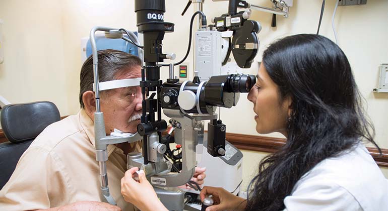
How is Retinal Detachment Treated?
Surgery, most of it done on an outpatient basis, is normally required. In the event of a retinal hole or tear that hasn’t progressed to an actual detachment, your ophthalmologist has several options, including the following:
- Laser surgery, which makes tiny welds around the hole, creating a seal that secures the retina in place. NYEE offers a full range of the latest tools for retinal laser photocoagulation along with wide-angle retinal imaging to give the surgeon the ability to perform the procedure in the most accurate and comfortable way possible.
- Cryopexy, which freezes the area around the hole by applying a probe to the outer surface of the eye directly over the hole or tear. This produces a seal that glues the retina to the wall of the eye.
In the case of a retinal detachment, several surgical procedures may be performed, including the following:
- Vitrectomy, where small needle-sized surgical instruments are inserted into the eye through the sclera (the white of the eye). These instruments remove the vitreous gel that fills the inside of the eye, along with any scar tissue pulling on the retina. Laser surgery or cryopexy is then applied to “weld” the retina in place. A supportive bubble of gas or silicone oil (like a cast on a broken limb) may be injected at the end of the surgery to temporarily replace the vitreous while the eye heals. The gas reabsorbs in the weeks following surgery, as the eye produces its own fluid to replace the excised vitreous gel. If silicone oil is used, an additional surgery will be necessary to remove it from the eye. NYEE’s retina surgeons have extensive experience performing minimally invasive, small gauge vitrectomies to minimize complications and promote quicker recovery.
- Scleral buckle, where the surgeon attaches a tiny synthetic band to the outside of the eyeball over the affected area. This surgical procedure indents the wall of the eye, gently supporting the damaged area of the retina that has produced the detachment. Scleral buckling usually includes laser treatment or cryopexy and may be performed in conjunction with vitrectomy.
When caught early, the vast majority of retinal detachments can be successfully treated with one of the above procedures, though additional procedures may sometimes be required in complicated cases. Early recognition of signs of visual disturbance is your best defense, especially if you are in a higher risk group.