Medical Photography and Imaging Service
The Department of Medical Photography/Imaging provides diagnostic ophthalmic services at the New York Eye and Ear Infirmary of Mount Sinai (NYEE) Retina Services Center. Highly specialized diagnostic imaging technologies and equipment are used to evaluate and document parts of the eye providing detailed information so that physicians can accurately diagnose conditions and develop effective treatment plans.
Your physician will need to complete a Medical Photography and Imaging Orders Form that indicates your specific service requirements prior to making an appointment. Please bring this form along with your insurance referral and/or pre-authorization (as required) on the day of your appointment.
NYEE is one of the most well equipped facilities in the Tri-state area with a wide range of state-of-the-art diagnostic equipment, allowing patients to get a complete diagnostic workup of their eyes in one location. We offer a wide range of tests, including the following.
Pentacam Corneal Topography
The Pentacam® HR is a high-resolution rotating Scheimpflug camera system for anterior segment analysis. It provides crisp images of the cornea, iris, and crystalline lens. It measures the anterior and posterior corneal topography and elevation, total corneal refractive power, corneal power distribution, automatic chamber angle measurement in 360°, chamber depth and volume, HWTW, and corneal and crystalline lens optical opacities.
Scanning Confocal Microscopy (Specular Microscopy)
The Nidek Confoscan 4 is the most advanced instrument of its kind. It is auto-aligning and is not affected by corneal haze and opacities. The one-of-a-kind Confoscan 4 also includes automatic cell-analysis software that detects the number of sides and area of each cell, as well as endothelial cell density, providing real-time in-vivo histology, and full cornea/endothelium/epithelium scans.
IOL Master
The Zeiss IOL Master provides non-contact axial length, K's, anterior chamber depth, and white to white measurements in a fast and reliable manner. Measurement values provide data for multi-formula and multi-lens calculations in a matter of seconds.
Slit Lamp Photography
With magnifications from 6x to 40x, slit-lamp digital imaging in the hands of our expert photographers provides superior documentation of anterior segment pathologies and anomalies.
B-Scan Ultrasound, UBM, and A-Scan
The 10MHz and 20MHz B-Probes provide high-resolution imaging of the retina and the orbit, with A-scan providing axial length measurements. Precise measurements of intraocular tumors are performed by our highly skilled and trained staff, under the guidance of one of New York's foremost ophthalmic oncologists.
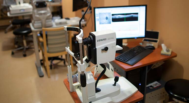
Optical Coherence Tomography (OCT)
Utilizing Heidelberg and Zeiss technology, OCT Spectral Domain Imaging has become the standard of care in ophthalmology. High definition cross sectional views of the retina, retinal nerve fiber layer, choroid (EDI), and anterior segment provide unsurpassed diagnostic imaging.
Fundus Photography
With recent technological advances in retinal imaging, traditional 20-, 30-, and 50-degree Fundus photography is still performed, as well as Optos wide field color imaging of the retina. Photographers are certified for most national studies by national reading centers.
Retinal Angiography
Fluorescein angiography of the retina and ICG angiography are performed by certified angiographers on either Topcon or Heidelberg systems, while the Optos Wide Field System performs strictly fluorescein angiography. In addition, auto fluorescence is performed on both Heidelberg and Optos systems.
Optical Coherence Tomography Angiography (OCTA)
Optical Coherence Tomography Angiography (OCTA) is a new, non-invasive imaging technique that uses light waves to take cross-section pictures of your retina. With OCTA, your ophthalmologist can see each of the retina's distinctive layers in order to isolate specific areas of interest. It has the ability to detect problems in the eye prior to any symptoms being present in the patient.
Microperimetry
Microperimetry is a technology that allows concurrent analysis of structural and functional aspects of the retina, and is one of several visual field tests used to create a retinal sensitivity map for patients who have lost the ability to fixate on an object. This is a powerful tool used to detect, describe, and follow pathologies affecting the macular area.