Conditions We Treat
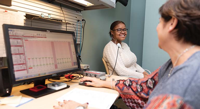
The specialists at the Ear Institute treat a wide range of conditions related to chronic ear disease, hearing loss, tumors, dizziness, and facial nerve disorders. Certain forms of hearing loss are caused by the blockage of sound along its pathway to the inner ear. This causes a conductive hearing loss. Depending upon the cause, some forms of conductive hearing loss can be surgically corrected. A complete examination by an otologist with a professional audiogram test will identify these conditions. Patients with asymmetrical neurosensory hearing loss, tinnitus, or dizziness should also see an otology-neurotology specialist with treatment options including medication, surgery and vestibular rehabilitation.
Our Ear Institute specialists have considerable skill and experience treating a wide range of conditions, including the following:
Hearing Disorders
- Acoustic Neuroma
- Acute Otitis Media or Ear Infections
- Auditory Neuropathy Spectrum Disorder (ANSD)
- Autoimmune Inner Ear Disease (AIED)
- Cerebrospinal Fluid Leak
- Cholesteatoma
- Dehiscent Superior Semicircular Canal Syndrome (DSSCS)
- Glomus Tumors
- Meniere's Disease
- Microtia
- Otosclerosis
- Perilymphatic Fistula
- Single-Sided Deafness (SSD)
- Temporal Bone Cancer
- Tinnitus
Balance Disorders
- Acoustic Neuroma
- Benign Paroxysmal Positional Vertigo (BPPV)
- Cervicogenic Dizziness
- Concussion
- Dehiscent Superior Semicircular Canal Syndrome (DSSCS)
- Imbalance of Aging
- Meniere’s Disease
- Migraine
- Perilymphatic Fistula
- Superior Canal Dehiscence (SCD)
- Vestibular Neuronitis and Labyrinthitis
Acoustic Neuroma
An acoustic neuroma, or a vestibular schwannoma, is a benign tumor that grows on the nerve responsible for hearing and balance in the inner ear, which sends information from the ear to the brain. Typically slow-growing, most acoustic neuromas are diagnosed between the ages of 30 and 60, and affect more women than men.
The most common symptom of an acoustic neuroma is hearing loss in the affected ear. This loss may be mild but is usually progressive. Other symptoms include a ringing noise (tinnitus) in the ear, or a sense of ear fullness. Many patients complain of vague imbalance or, occasionally, vertigo (the false sensation of moving). When an acoustic neuroma is large and pushes against the brain, it can cause headache and facial numbness and weakness.
Treatment of an acoustic neuroma depends upon its size, rate of growth, and location, as well as the amount of hearing loss and the age of the patient. Common approaches to treatment are careful observation, surgical removal, and focused radiation therapy (radiosurgery).
Acute Otitis Media or Ear Infections
A bacterial or viral infection of the middle ear is known as acute otitis media (AOM)—the most common reason for childhood visits to the pediatrician. Because of the inflammation and build-up of fluids, AOM is often painful, especially for young children. Some youngsters may run a fever and have diarrhea, irritability, and feel generally sick. Children are more likely than adults to get ear infections.
Acute otitis media may heal on its own, requiring only pain medication. When the condition is more serious or involves an infant, antibiotics such as amoxicillin are an effective therapy.
If non-infected fluid collects in the middle ear as the result of a cold, sore throat, or upper respiratory infection, it is known as otitis media with effusion (OME). Symptoms may include hearing difficulties and a feeling of fullness in the ear. OME is most common in children between six months and three years of age. In most cases, treatment for OME is not required since the fluid accumulation resolves on its own. If the condition persists for several months, your child’s pediatrician or ENT (otolaryngologist) may recommend a minor surgical procedure known as myringotomy, in which ventilation tubes are placed in a small opening in the eardrum to drain the fluid and relieve the pressure. Hearing is soon restored.
Auditory Neuropathy Spectrum Disorder (ANSD)
Auditory Neuropathy Spectrum Disorder (ANSD) results from the abnormal transmission of sound from the hair cells of the inner ear to the hearing center of the brain. Children who are born prematurely or with hypoxia (low oxygen) or elevated bilirubin at birth are at greater risk of this condition. A family history of the disorder can also play a role.
ANSD occurs in approximately 10 percent of people with profound hearing loss, and is being diagnosed more frequently in children due to newborn neonatal screening. A consequence of this disorder is poor word understanding relative to the level of hearing sound.
There is no cure for ANSD. Early diagnosis and treatment, however, can result in children developing strong language and communication skills with the help of assistive listening devices, such as hearing aids and cochlear implants.
Autoimmune Inner Ear Disease (AIED)
Autoimmune Inner Ear Disease (AIED) is a rare disease that occurs when the body's immune system attacks cells in the inner ear, which it mistakes for a virus or bacteria. Symptoms of AIED are sudden hearing loss in one ear progressing rapidly over weeks or months to the other ear. Patients may also experience a fullness in the ear accompanied by a ringing, hissing, or roaring sound. About half of patients with AIED experience balance problems in the form of dizziness and unsteadiness.
AIED is a very difficult inner ear problem to diagnose. Many patients with AIED respond to an initial treatment of steroids and may be helped by other, newer medications designed to target various inflammatory mediators. Patients with AIED often benefit from the care and input of a rheumatologist familiar with AIED.
Balance Disorders
As a teaching center with a high patient volume, the specialists at the Ear Institute of New York Eye and Ear Infirmary of Mount Sinai are experienced in treating all types of vestibular dysfunction and offer a hands-on approach to a number of balance disorders, including:
- Acoustic neuroma is a benign tumor that grows on the nerve affecting hearing and balance within the inner ear—the same nerve that sends information from the ear to the brain. Vestibular rehabilitation may be helpful in addressing balance deficits that result from this compromised nerve.
- Benign paroxysmal positional vertigo (BPPV) is the most common cause of dizziness and vertigo in adults. BPPV occurs when tiny calcium crystals break off and collect as loose debris (otoconia) within the inner ear. Our therapists are specially trained to use a series of customized head exercises to relocate the otoconia back to its proper position.
- Cervicogenic dizziness results from whiplash injuries occurring with head traumas that involve injures to the bone, muscles, or nerves of the cervical spine; herniated cervical spine discs; muscle spasms in the neck; or arthritis. A combination of orthopedic surgery and vestibular rehabilitation can address the underlying structural issues, as well as the dizziness and disorientation this condition causes.
- Concussion is caused by a jolt or blow to the head. Vestibular rehabilitation can address dizziness, imbalance, or vertigo following a brain injury resulting in a concussion syndrome or the inner ear injury.
- Dehiscent superior semicircular canal syndrome (DSSCS) is caused by an abnormal opening (dehiscence) in the bone overlaying the superior semicircular canal of the inner ear. Though this condition is often managed surgically, patients may undergo vestibular rehabilitation to reduce the dizziness and vertigo resulting from this rare inner ear condition.
- Imbalance of aging is the age-related impairment of the major senses we need to maintain our balance. Vestibular rehabilitation directly addresses this vestibular deficit, and teaches individuals strategies to mitigate the loss of other senses, with an emphasis on safety, independence, and the prevention of falls.
- Meniere’s disease is caused by the abnormal buildup of fluid in the inner ear. Vestibular rehabilitation may help to improve balance in mid-to-late-stage disease through exercises and activities that involve head motion.
- Migraine can result in motion sensitivity (vertigo) that impairs a person’s ability to function normally. Vestibular rehabilitation, in conjunction with medical management of the migraine, can help reduce motion sensitivity and restore some level of function.
- Perilymphatic fistula is caused by the leakage of fluid through a torn membrane that separates the inner ear from the middle ear. While this condition is usually managed surgically through membrane repair, some patients may benefit from post-surgical vestibular therapy to manage any residual symptoms of dizziness and imbalance.
- Superior canal dehiscence (SCD) is managed surgically, but patients are often asked to undergo a trial of Vestibular Rehabilitation as a conservative approach to try to reduce the patient’s dizziness. Patients may also benefit from post-surgical therapy, should they have any residual symptoms of dizziness and imbalance following the procedure.
- Vestibular neuronitis and labyrinthitis are inflammations caused by viral infection that can cause damage to hearing and vestibular function (labyrinthitis) or damage to vestibular function alone (vestibular neuronitis). Vestibular rehabilitation allows patients to explore and recalibrate their balance, with or without head movement.
Cerebrospinal Fluid Leak
This often under-diagnosed but serious condition is caused by a hole in the bone separating the brain from the ear. As a result, cerebrospinal fluid drains from the brain into the ear canal. Patients will complain of clear, thin fluid in the ear, which is constantly leaking, or a funny taste in their throat because they are swallowing the fluid.
The hole can occur as the result of trauma, infection, a past surgical issue, or spontaneously. Obesity may also play a role; as the pressure inside the skull increases, and the brain begins to pulsate on the bone, it may eventually break and leak. Prompt diagnosis and treatment is needed in order to avoid possible complications, including meningitis or a brain abscess.
The location of the hole will determine the proper surgical treatment. In many cases a multispecialty team consisting of a neurosurgeon and an otologist-neurotologist will patch the hole both above and below the ear to stop the leakage.
Cholesteatoma
Cholesteatoma is an abnormal cyst filled with shed skin. There are two types of cholesteatoma:
- Congenital cholesteatoma results from skin remnants that fail to migrate from the middle ear to the outer ear during fetal development. This condition appears in children as an asymptomatic white mass behind the ear drum (tympanic membrane).
- Acquired cholesteatoma results from untreated ear fluid (middle ear effusion), recurrent ear infections, or an untreated hole in the ear drum. Shed dead skin in the middle of a cholesteatoma cyst often becomes chronically infected, and if untreated can be very destructive to the bone of the ear.
Cholesteatoma can cause hearing loss, dizziness, and rarely, a weakness of the muscles of the face. It can also present as a chronic infection of the ear. The size of a cholesteatoma is usually determined through computed tomography (CT). Cholesteatoma is treated surgically in the majority of cases.
Dehiscent Superior Semicircular Canal Syndrome (DSSCS)
Dehiscent Superior Semicircular Canal Syndrome (DSSCS) results from an abnormal opening (dehiscence) in the bone overlaying the superior semicircular canal of the inner ear. Patients with this syndrome can experience dizziness and vertigo (a feeling of motion and loss of balance), often precipitated by loud noise or sudden motions that alter the inner ear pressure, such as coughing, sneezing, or lifting heavy objects. Hearing loss is also common.
DSSCS is confirmed by specialized balance testing called vestibular evoked myogenic potential (VEMP), as well as CT scans of the temporal bones of the inner ear. Many patients with the disorder are able to tolerate their symptoms by reducing stimuli, such as loud noises. For others whose quality of life is impacted, surgical repair of the dehiscence can be effective.
Glomus Tumors
Glomus tumors, or paragangliomas, are usually benign, slow-growing, but highly vascular tumors. When the tumor is present inside the middle ear, the growth can block sound, causing conductive hearing loss. Patients often complain of hearing a pulsating sound (a form of tinnitus) due to the high blood flow within the tumor. If the patient’s inner ear is affected, they may experience sensorineural hearing loss or vertigo.
A CT or MRI scan along with an examination will help to diagnose the nature of the growth and its location. The choices of management include observation, radiation therapy, or surgery.
Meniere's Disease
Meniere's disease results from an abnormal buildup of fluid in the inner ear, presenting as a feeling of fullness or pressure in the ear, ringing (tinnitus), hearing loss, and vertigo. These symptoms may occur suddenly, last from minutes to hours, resolve spontaneously, and recur at irregular intervals. Meniere’s disease is a chronic condition that usually affects one ear. Over time, it may result in permanent hearing loss, while vertigo can increase your chances of falls and accidents while driving.
Although no cure exists for Meniere’s disease, your ENT specialist may prescribe medicines to alleviate the vertigo. The use of diuretics, which reduce fluid retention, may help control the symptoms of the disorder. Vestibular rehabilitation may help to improve your balance in mid-to-late-stage disease through exercises and activities that involve head motion. In the most severe cases of the disease, surgery or injections of medicine into the middle ear are treatment options.
Otonomy Meniere's Disease Trial
Microtia and Atresia
Microtia is an uncommon disorder in which one or both of a baby’s external ears are malformed. This congenital condition can be mild or so severe that the ear is missing entirely. Microtia frequently occurs with aural atresia, the absence of normal ear components (such as the tympanic membrane or ear canal), which causes moderate to severe hearing loss.
Following an evaluation, treatment at our Microtia and Atresia Program involves reconstruction surgery, and possible additional procedures, which may involve, opening the ear canal, bone-anchored hearing aid procedures, or an external prosthesis.
After surgery, our specialists will personalize a treatment plan (which may include hearing aids or cochlear implants) with the goal of achieving the best level of hearing possible.
Otosclerosis
Otosclerosis is a disease of one of the tiny, innermost bones (the stapes) of the inner ear that vibrates and transmits sound from the middle to the inner ear, enabling us to hear. This often genetically-linked disease usually begins in the late twenties or thirties, is more common in women than men, and is bilateral (affects both ears) in half of patients. Otosclerosis causes gradual hearing loss and may also be associated with tinnitus and ear fullness or pressure.
If hearing loss is mild or unilateral, otosclerosis may require only careful observation. More serious cases may need amplification in the form of a hearing aid. Another option is surgical removal of the stapes bone and its replacement with a prosthesis (a procedure known as a stapedectomy).
Perilymphatic Fistula
Perilymphatic fistula is a tear in one or both of the thin membranes that separate the air-filled middle ear from the fluid-filled perilymphatic space of the inner ear, resulting in leakage of microscopic amounts of fluid into the middle ear. This condition is due to sudden changes in middle ear pressure, which directly affect the inner ear. The most common causes include head trauma, ear trauma from objects perforating the eardrum, and sudden descent of an airplane or the aftermath of a scuba dive.
Perilymphatic fistula is extremely difficult to diagnose. Treatment may involve surgical repair of the tear in the membrane.
Tinnitus (Ringing in the Ears)
Tinnitus is characterized by ringing sounds in the ear. It can be triggered by a wide range of underlying conditions, including exposure to loud noise, middle ear injuries, a hole in the eardrum, earwax, cardiovascular disorders, allergies, high or low blood pressure, a tumor, diabetes, injuries to the head or neck, or certain medicines. Tinnitus may be periodic or continuous, and it may vary in pitch from a low roar to a high squeal or whine. It may be present in one or both ears. Millions of people with tinnitus are so severely afflicted they cannot lead normal lives.
If your tinnitus is bothersome, it is important to see an ear specialist (otolaryngologist), who will attempt to determine the cause and decide on the best treatment plan. In some cases, noise suppression is an effective strategy. This may include masking devices that are worn in the ear like a hearing aid and produce a low-level white noise that muffles the tinnitus. Some physicians suggest listening to FM music at low volume, or white noise machines with pillow speakers to help you sleep. Concentration and relaxation exercises may also help to reduce the intensity of the problem in some patients by controlling muscle groups and circulation throughout the body. Occasionally, medicines may help reduce the severity of the symptoms.
Tinnitus in Children
Tinnitus in children is rare, though when it occurs the child will not typically complain about it since they consider the noise in their ear to be normal. Ongoing tinnitus can be annoying and distracting, however, and in severe cases can cause psychological distress and interfere with your child’s ability to lead a normal life. The good news is most children with tinnitus seem to eventually outgrow it.
If you suspect your child has tinnitus, make an appointment with your family physician or pediatrician. In most cases, no underlying problem will be found, and simply having the condition and ways to manage it explained to the child by a medical professional will help. In more serious cases, a referral to an otolaryngologist (ear, nose, and throat specialist) may be necessary. Depending on the nature of the tinnitus, the doctor may order comprehensive hearing tests.